Chest X-ray
A chest X-ray gives the cardiologist a picture of the size and shape of your child’s heart and indicates the presence or absence of fluid in the lungs. Different views are common, and may include the front and side of the child, standing or lying.
Electrocardiogram
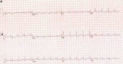
An electrocardiogram (EKG) measures electrical activity in the heart, the heart’s rate and rhythm, damage or thickening of the heart muscle and any unusual events in the conduction system. The test takes about 15 minutes and requires only that several wired patches be placed on the chest, arms and legs. For additional information, please read about our Heart Station.
Echocardiography
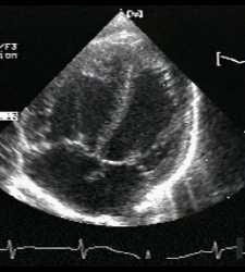
Echocardiography enables the cardiologist to see details of the heart structure from outside the body and helps determine the need for further evaluation or treatment. Echocardiography uses high-frequency sound waves – also know as ultrasound – to form an image of heart tissues and blood flow. All body tissues and fluids absorb or reflect these sound waves in specific ways and create an “echo,” which can be measured. The echocardiography technician creates an image of the heart by measuring the waves’ reflections. The test will last about 30 to 45 minutes. During this test, all the child will feel is the transducer touching his or her chest. A transducer is a flat-bottomed instrument, about six-inches long, that moves smoothly along the chest. A clear gel will be applied to the chest to help the transducer move smoothly. Repetitive pulses of ultrasound waves, silent to our ears, are delivered to the heart. Between these pulses, the returning echoes are gathered and recorded. Sedation is sometime necessary to ensure good pictures. Your child may be given something to drink or nasal spray that will help him or her to go into a light sleep. For additional information, please read about our Heart Station.
Holter monitor
A Holter monitor measures heart rate and rhythm for a 12 to 24-hour period. It is commonly worn by a patient before and after cardiac surgery, so the doctor can compare before and after information on the heart’s electrical activity. A Holter monitor is a box carried on a shoulder strap or belt with wires that are painlessly connected to the chest with patches. The patient’s nurse or family keeps a diary of the child’s activities, such as walking, eating and crying, and the time they occur. The doctor uses this information to see how the activities affect heart rate and rhythms. For additional information, please read about our Heart Station.
Cardiac catheterization and angiography
The day before the cardiac catheterization, your child will receive precatheterization testing, which includes cardiac tests, and chest X-ray. This testing begins at the Heart Station. A Cardiologist will meet with you to review your child’s medical history, explain the cardiac catheterization procedure, and obtain a signed consent form. You also will be told when your child should not have anything more to eat or drink. On the morning of the cardiac catheterization, you should go to Admitting at your scheduled time.
When your child is called for the cardiac catheterization, the nurse will ask him or her to use the bathroom. One hour prior to the procedure, a premedication cream will be placed on both groin areas to numb the areas. The child also may be given a premedication to drink that takes him or her drowsy. You may stay with and hold your child until he or she is ready to enter the cardiac cath lab. At that time, you will be asked to return to your child’s room. The procedure lasts approximately three hours. If you leave your child’s room, let the nurse know where you will be. As soon as the procedure is completed, your child returns to his or her room, and a cardiologist will discuss the preliminary findings with you. A thick pressure dressing of elastic tape and gauze sponges will cover the catheter site. Your child may be sleepy for a while from the sedative used during the procedure. To help prevent bleeding, the child should lay flat for several hours. Your child may receive fluids when he or she awakes, and diet will be advanced slowly as tolerated.
If surgery is not planned for the following day, your child may be dischargedthe same day as the catheterization or the next morning. Before discharge, a cardiologist sees your child and determines whether it is appropriate to leave. You will receive discharge instructions and supplies needed for wound care.
Electrophysiology study
An electrophysiology study (EPS) measures the heart’s electrical system to help assess arrhythmias, which are abnormal heart rhythms. An EPS is similar to a cardiac catheterization. It is performed by inserting catheters into veins and advancing these into the heart. The movement and location of the catheter are shown to the doctor by X-rays displayed on a television screen. Electrical wires with sensors on the ends are passed through veins to certain areas of the heart. The wires pace the heart at different rates so the cardiologist can analyze arrhythmias. The procedure also allows the cardiologist to map the heart’s electrical conduction system from inside and look for places in the heart that are over-sensitive or under-sensitive to electrical impulses. If an over-sensitive area is found to cause the arrhythmia, medical or surgical treatment may be needed to eliminate the arrhythmia. If an under-sensitive or blocked pathway is found, the surgeon may prescribe medication or a pacemaker to correct the heartbeat.
Magnetic resonance imaging
Magnetic resonance imaging (MRI) provides a detailed view of the heart. The MRI equipment creates a large magnetic field that causes atoms in the body’s tissue to emit radio waves that are received by an antenna. A computer converts the signals into visible images of the heart and arteries. The child will lie on a sliding table that goes inside a large tube. Except for EKG leads attached to the child’s chest with adhesive, nothing touches the child, and the child will not feel any sensations. The exam lasts about one hour.